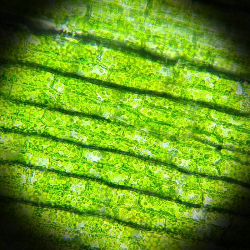
This cell was taken from waterweed on 29.9.25. You can't see the nucleus, our teacher told us it's because the chloroplast covers it. It's hard to see the cell walls, but they are kind of outlined by clusters of chloroplast. All of the green specks are the chloroplast.
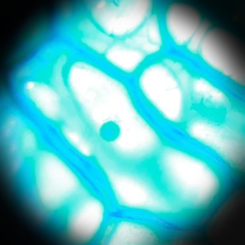
This cell was taken from an onion (white) on 29.9.25. It was colored with methylene blue, which makes the cell wall and nucleus easier to see. I don't think I have to point out where these are.
.png)
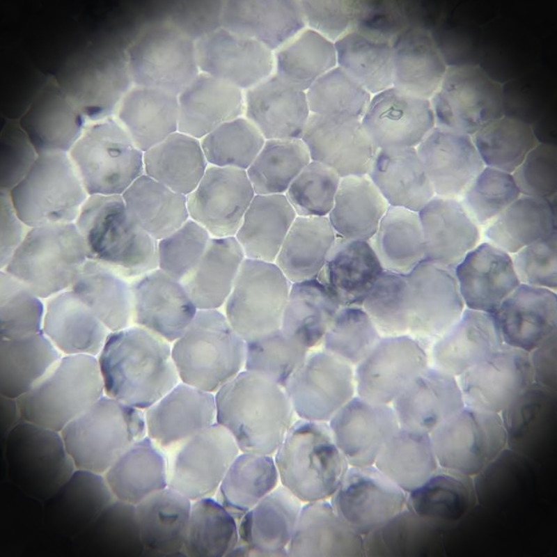
This cell was taken from my cheek mucous membrane on 29.9.25. It was also colored with methylene blue. I am an animal, so my cells don't have any walls, and in the image you can't see the celmembrane, since these usually aren't shown on light microscopes. You can see the nucleus, though.
This cell was taken from a petal - I'm not sure from what flower it came - on 29.9.25. I'm pretty sure you can see the cell walls, but you can't see much else, which was kind of strange. On other things I saw - such as the waterweedcells - you couldn't see the nucleus because it was covered by the plastids. But you can't see the chromoplast here either, which I had expected to see, too. I also looked at a tomato cell (not pictured) and the chromoplast was hard to see, so maybe they were present here.
.png)
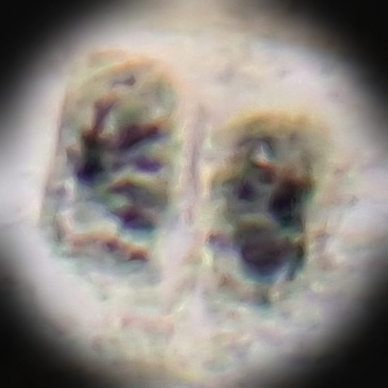
This cell is also from my own cheek mucous membrane, taken on 29.9.25. Colored with methylene blue. I made this one first, and if you can see the cell isn't very "good". None of the cells in this preperation were "good", so I had to make the other ones. When I first saw this, I honestly thought I had some crazy ilness or something, but my teacher told me that, because my cells have no cell walls, the cells just managed to merge together - the preperation was difficult to make, so that might have caused this. In the picture of the other cell, you can also see a little fold on the left underside. In this picture, you can see multiple cell nuclei, I think 2-3.
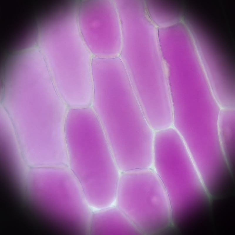
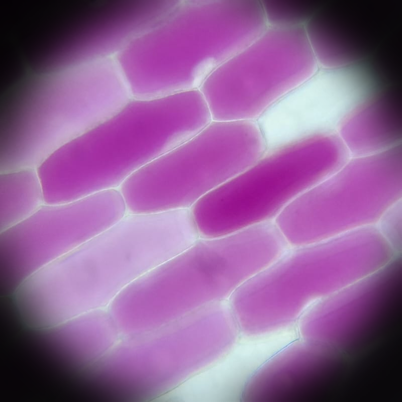
This cell was taken from an onion (purple/red) on 20.10.25. It was put into a solution of 0% Potassium Nitrate (KNO3). The KNO3 was a hypotonic solution, meaning that it had a lower concentration of dissolved particles outside of the cell compared to inside the cell. Osmosis causes water to move from a lower concentration of disolved particles to a higher concentration. So, water from outside of the cell was entering the cell, causing it to become turgid (which, plants prefer to be a little bit turgid, since they get strength from the water).
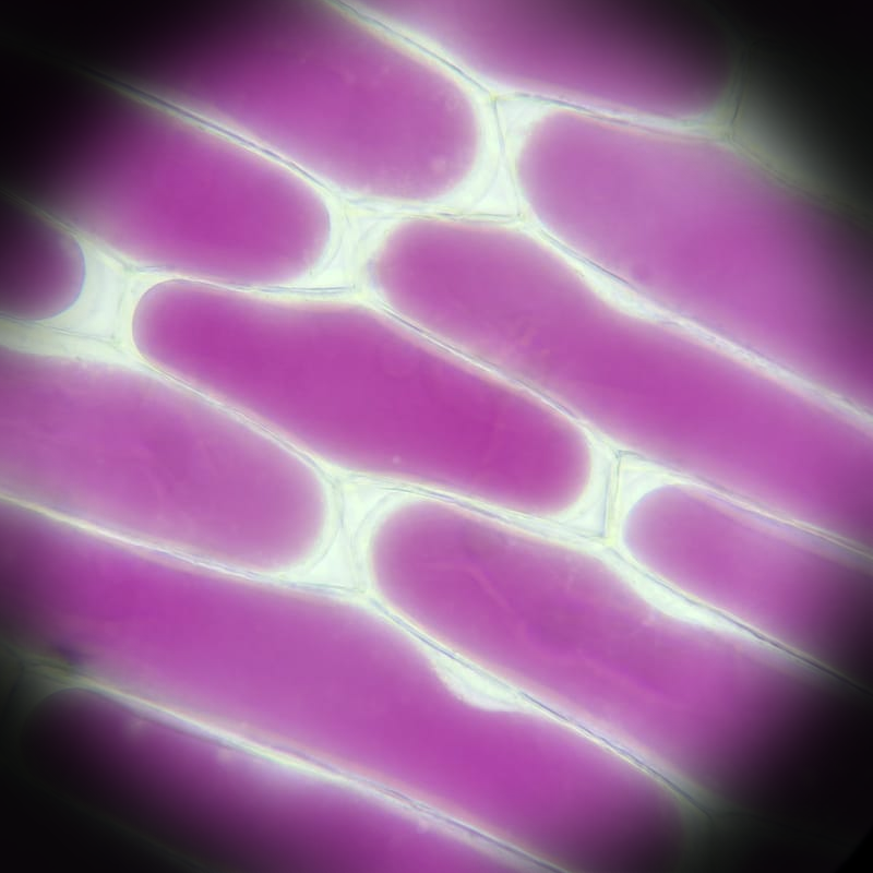
This cell was taken from an onion (purple/red) on 20.10.25. It was put into a solution of 2% (KNO3). The KNO3 was a hypotonic solution, so this cell was also turgid.
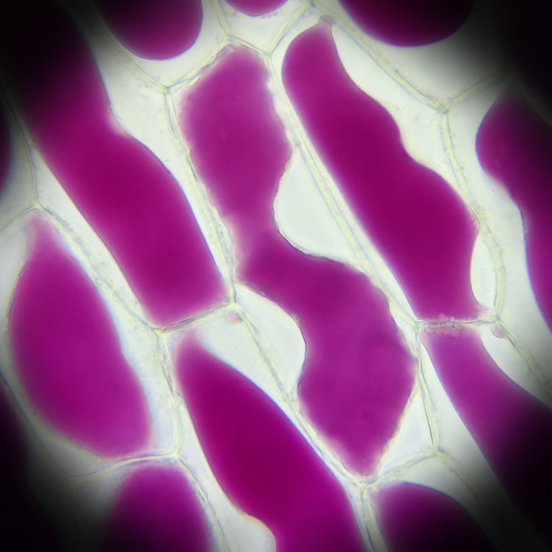
This cell was taken from an onion (purple/red) on 20.10.25. It was put into a solution of 4% (KNO3). The KNO3 was a isotonic solution, meaning that it had an equal concentration of dissolved particles outside of the cell compared to the inside of the cell. In this situation, water leaves and enters equally, too. So this is how a cell would "normally" look (this isn't ideal for a plant cell, though).
This cell was taken from an onion (purple/red) on 20.10.25. It was put into a solution of 6% (KNO3). The KNO3 was a hypertonic solution, meaning that it had a higher concentration of dissolved particles outside the cell compared to the inside of the cell. So, water from inside the cell was exiting the cell, causing it to become plasmolyzed.
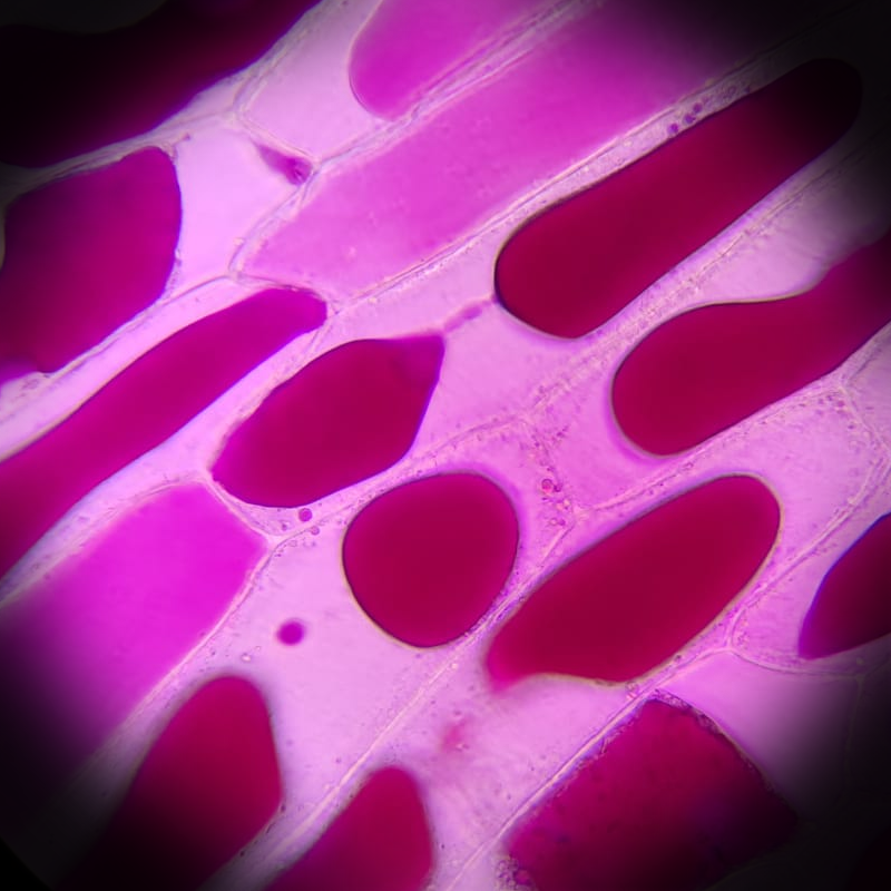
This cell was taken from an onion (purple/red) on 20.10.25. It was put into a solution of 10% (KNO3). The KNO3 was a hypertonic solution, so this cell was also plasmolyzed. (This image looks so pink because a lot of the color from the onion leaked out onto the slide, coloring the KNO3).
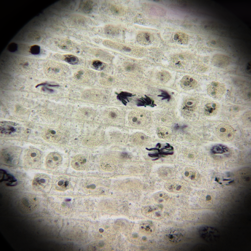
These are onion cells, I'm not sure when the slide was made (they were prepared for us), but I looked at it on 17.11.25. It was looked at with the light microscope at a magnification of 100x10 (all of my other pictures are taken at 40x10) and they were looked at with immersion oil. In this picture you can see the stages of mitosis in the onion cell. Not all are visible, only anaphase and prophase (to my knowledge).
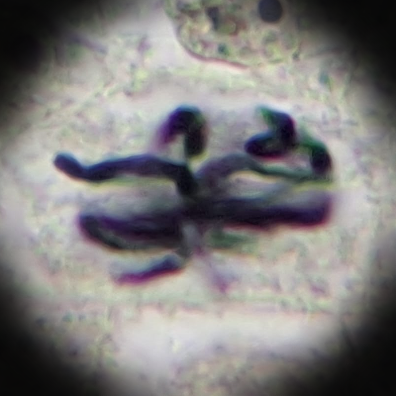
A close up of prophase (might be blurry, since it's cropped from a larger microscope image). Mitosis happens in these steps: interphase (not shown, since it's harder to see with a light microscope) -> prophase -> metaphase -> anaphase -> telophase -> cytokenisis (also not shown). My textbook describes prophase like this (translated from Dutch to English): "The chromosomes become shorter and thicker (spiralization), the nuclear membrane disappears, spindle emerges from the centrioles."
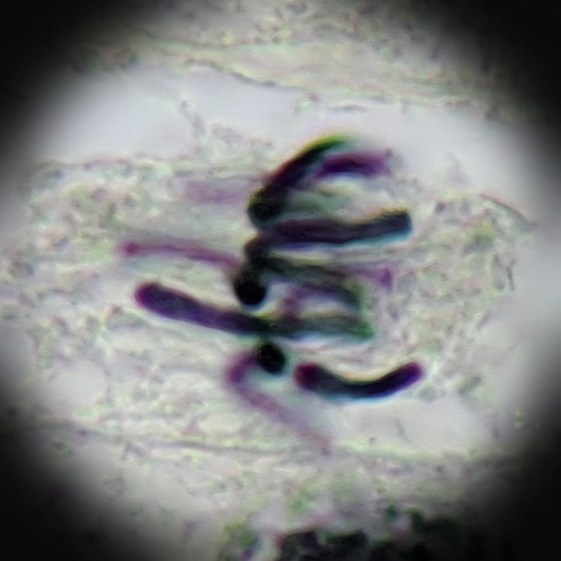
A close up of metaphase (might be blurry, since it's cropped from a larger microscope image). My textbook describes metaphase like this (translated from Dutch to English): "The chromatids arrange themselves in the equatorial plane between the two poles." So basically you can recognize metaphase when you notice a line of chromatids in the middle of your cell (it's not always 100% perfect).
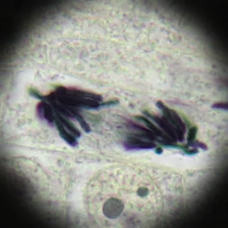
A close up of anaphase (might be blurry, since it's cropped from a larger microscope image). My textbook describes anaphase like this (translated from Dutch to English): "Chromatids are split down the middle, the daughter-chromatids go to the poles." Also important to note that the "poles" (spindles) are located on opposite sides of the cells.
A close up of telophase (might be blurry, since it's cropped from a larger microscope image). My textbook describes telophase like this (translated from Dutch to English): "New nuclear membranes and cell membrames form around the daughter-chromosomes". I want to note that chromosomes and chromatids aren't the same thing, but the explanation is way too long (even when simplified), so I do recommend looking it up if you're interested!